Horse Leg Bones Diagram / Forever Horses Anatomy Of The Equine Hindleg - When the skeletal structure is properly proportioned the joints work smoothly.
Horse Leg Bones Diagram / Forever Horses Anatomy Of The Equine Hindleg - When the skeletal structure is properly proportioned the joints work smoothly.. Horse anatomy diagrams legs via. Look at the diagram on the previous page of the front legs and toes (hooves) of some of these horse fossils. In this picture it shows the muscles that are closest to the surface of the skin, making them superficial. This is because there are many layers of muscles. Start studying leg joints and bones of the horse.
Principles of bone development in horses. At this stage of life, even with this exceptionally early development, horses have only 17% of their mature bone mineral content. When the skeletal structure is properly proportioned the joints work smoothly. Diagram of horse body parts. The legs of a horse are made up of a system of muscles, tendons, ligaments, and connective tissue.
The Horse S Skeleton Hind Limbs Youtube from i.ytimg.com This is because there are many layers of muscles. Their leg bones are proportioned differently from those of a human. It covers the front and sides of the third phalanx, or coffin bone. Similarly, the hock contains the bones equivalent to those in the human ankle and heel. Horse anatomy diagrams legs via. If the angle at witch these bones are working is compromised, the joint becomes unevenly stressed and injury to the tendons and ligaments. Performance horses tend to suffer from this degenerative disease. This is supposed to demonstrate a change from browsing on bushes to grazing on grass.
The horse leg anatomy in the rear includes the bones of the pelvis (the ilium, ischium and pubic bones), femur, tibia, fibula, metatarsus and the phalanxes.
This system works together to support horses weight when it stands up also works to diminish compression during movement which helps to horse to avoid injury to their limbs. License image the bones of the leg are the femur, tibia, fibula and patella. Horse anatomy diagrams legs via. These bones are called the carpal bones, except for the 7th bone which is referred to as the accessory carpal bone. The horses legs and hooves are also unique, interesting structures. Proper hoof care, good exercise management. The blood supply to and/or from the navicular bone is disrupted. Few animals are as precocious as the horse. In this picture it shows the muscles that are closest to the surface of the skin, making them superficial. How many front toes did the oldest horse have? It covers the front and sides of the third phalanx, or coffin bone. The ulna and the radius. Flat footed horses (ie, those whose pedal bones lie flat instead of being tilted slightly on their nose) often have severely atrophied digital cushions.
The ulna and the radius. Few animals are as precocious as the horse. This diagram shows the superficial layer of the tissue. The horses legs and hooves are also unique, interesting structures. Similarly, the hock contains the bones equivalent to those in the human ankle and heel.
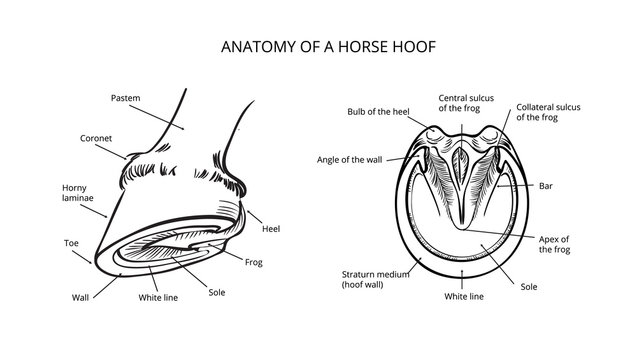
256 Best Horse Hoof Anatomy Images Stock Photos Vectors Adobe Stock from t4.ftcdn.net Human anatomy muscles coloring pages printable via. This is because there are many layers of muscles. We make sure to keep the original photos without changing. Both pelvic and thoracic limbs contain the same number of bones, 20 bones per limb. Flat footed horses (ie, those whose pedal bones lie flat instead of being tilted slightly on their nose) often have severely atrophied digital cushions. The wall is made up of the toe (front), quarters (sides) and heel.when the foot is lifted off the ground, the sole and frog are visible, as well as the bars of the wall and the collateral. The ideal horse has legs which are straight, correctly set and symmetrical. The bones of the horse skeleton are held together with ligaments, tendons and muscles.
The power propulsion system and major defensive tool, a horse's rear.
Horse vs human heather smith thomas made a beautiful comparison, in her book the horse conformation handbook, between the anatomy of the horse's lower leg and that of the human hand. The ideal horse has legs which are straight, correctly set and symmetrical. Horse leg structure there are also many fossil remains of horse leg bones. The top part of the hind limbs consists of three fused bones, called the ileum, ischium, and pubis. Long bones are found in the arms (humerus, ulna, radius) and legs (femur, tibia, fibula), as well as in. How many front toes did the oldest horse have? In this image, you will find second (medial) and 27.05.2019 · diagram of horse leg posted on may 27, 2019 by admin a horse has many bones in its legs horse leg diagram horse leg anatomy diagram. This is because there are many layers of muscles. Look at the diagram on the previous page of the front legs and toes (hooves) of some of these horse fossils. Horse stifle joint anatomy via. Flat footed horses (ie, those whose pedal bones lie flat instead of being tilted slightly on their nose) often have severely atrophied digital cushions. It is likely that abnormal biomechanical stresses are the basis for the disease. The wall is simply that part of the hoof that is visible when the horse is standing.
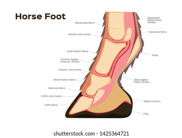
The eohippus horses part b. The bones of the leg are the femur, tibia, fibula and patella. Long bones are found in the arms (humerus, ulna, radius) and legs (femur, tibia, fibula), as well as in. The bones that make up the lower leg are the cannon bone, splint bones, long pastern, short pastern, pedal bone and navicular bone. It also includes the joints of the hip, stifle, hock, fetlock, pastern, and coffin.
Horse Anatomy Hd Stock Images Shutterstock from image.shutterstock.com These bones are called the carpal bones, except for the 7th bone which is referred to as the accessory carpal bone. Similarly, the hock contains the bones equivalent to those in the human ankle and heel. If the angle at witch these bones are working is compromised, the joint becomes unevenly stressed and injury to the tendons and ligaments. Health diagram bone skeleton leg knee science anchor chart human human body. Look at the diagram on the previous page of the front legs and toes (hooves) of some of these horse fossils. The top part of the hind limbs consists of three fused bones, called the ileum, ischium, and pubis. In our website, we are persons who very treasure creativity from every one, with no exception. There are many possible diagrams of the anatomy of horse tissues.
The horse's back leg is homologous to a human's leg, too, but we are going to focus on the foreleg and arm as our example for this blog.) beneath (or outward from) the humerus there are two bones, side by side:
Horse rear legs the horse leg anatomy in the rear includes the bones of the pelvis (the ilium, ischium and pubic bones), femur, tibia, fibula, metatarsus and the phalanxes. Similarly, the hock contains the bones equivalent to those in the human ankle and heel. Flat footed horses (ie, those whose pedal bones lie flat instead of being tilted slightly on their nose) often have severely atrophied digital cushions. Few animals are as precocious as the horse. Health diagram bone skeleton leg knee science anchor chart human human body. The top part of the hind limbs consists of three fused bones, called the ileum, ischium, and pubis. A corium is a vascular structure which manufactures hoof horn. There are many possible diagrams of the anatomy of horse tissues. The bones that make up the lower leg are the cannon bone, splint bones, long pastern, short pastern, pedal bone and navicular bone. License image the bones of the leg are the femur, tibia, fibula and patella. The pelvic limb typically contains 19 bones, while the thoracic limb contains 20 bones. Six small bones make up this joint, and it is often the site of strain and wear and a common location for arthritis. » science abc from www.scienceabc.com 13.05.2021 · horse leg bone diagram :
Lower extremity arterial duplex via leg bones diagram. Finally, there is the large modern horse, equus, with only one toe, while all that is left of the other two are 'vestigial' splint bones.


0 Komentar